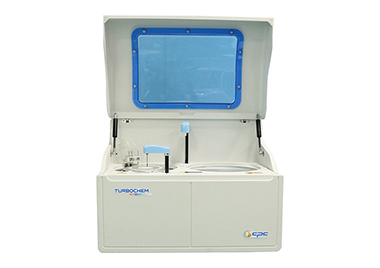
New Models
CPC Diagnostics

The fact that the immune system can target virtually any structure within the central or peripheral nervous structure has made neuroimmunology a rapidly evolving specialty. These neurological conditions manifest as disorders previously thought to be independent and unrelated. The spectrum of autoimmune neurological syndromes has expanded substantially in the last fifteen years due to the discovery of novel anti-neuronal antibodies. Discovery of new markers enables better diagnosis. In this line comes the novel marker for Neuromyelitis Optica – MOG (myelin oligodendrocyte glycoprotein). MOG is expressed exclusively in the central nervous system (CNS) on the outer layer of the myelin sheath and the plasma membrane of oligodendrocytes. Though the 28 kDa protein comprises only a fraction of the myelin sheath (<1%), it is an important surface marker which is involved in the myelination of CNS nerves. NMO and NMOSD are immune-mediated chronic inflammatory disorders of the central nervous system characterized by severe demyelination and axonal damage, targeting predominantly optic nerves and spinal cord. These rare neuroinflammatory diseases differ typically in the age of onset, disease severity, clinical course, neuroradiological and/or pathological characteristics, and changes in cerebrospinal fluid (CSF). Autoantibodies against aquaporin-4 (AQP-4), a water channel protein expressed on astrocytes, are an established biomarker for the disease. They can be detected in 60 to 90 percent of patients fulfilling the clinical diagnostic criteria for NMO. The detection of autoantibodies against AQP-4 (also termed as NMO IgG) is specific for NMO and also for recurrent opticus neuritides (ON) and longitudinal extensive transverse myelitis (LETM). The determination of autoantibodies against MOG is especially relevant for the diagnosis of acquired demyelinating diseases of the CNS, particularly in children and NMO. Double positivity of Aquaporin and MOG is rare. The diagnosis of demyelination, associated with AQP-4 or MOG-antibodies, is decisive for therapeutic measures and has a significant predictive value. In NMOSD, AQP-4 antibodies are detected in 64.7 percent of cases, while anti-MOG autoantibodies are only found in 7.4 percent. Patients with seropositivity for MOG and negative for AQP-4 are more likely to have ON and LETM and often exhibit a monophasic course. These patients have a good prognosis, connected with less recurrences than patients with a positive anti-AQP-4 serological result or double-negative patients (AQP-4 and MOG seronegativity). The anti-MOG IIFT determines autoantibodies against myelin-oligodendrocyte glycoprotein. This specific and sensitive test is based on transfected cells expressing the full-length protein. Recent studies show that 30–40 percent of pediatric patients with inflammatory demyelinating diseases of the CNS exhibit autoantibodies against MOG at disease onset. The antibody determination with Euroimmun IIFT enables early delimitation of neuromyelitis optica spectrum disorders (NMOSD) for important differential diagnoses, such as multiple sclerosis (MS). Multiple sclerosis was traditionally considered as a variant of NMO, but now it is recognized as a distinct clinical entity based on its unique immunological features. Diagnostic delimitation of acute demyelinating syndrome from multiple sclerosis (MS) is particularly important, since treatment for MS and NMO/NMOSD are very much different. Euroimmun serological tests for neurology can be performed in routine diagnostic laboratories. Neurologists can request the analyses from their usual laboratory.


You must be logged in to post a comment Login